Prices plus VAT plus shipping costs
Ready to ship today,
Delivery time appr. 5-8 days
- Order number: 7TM0358N-RM
- Content: 100 µl
- Host: Rabbit
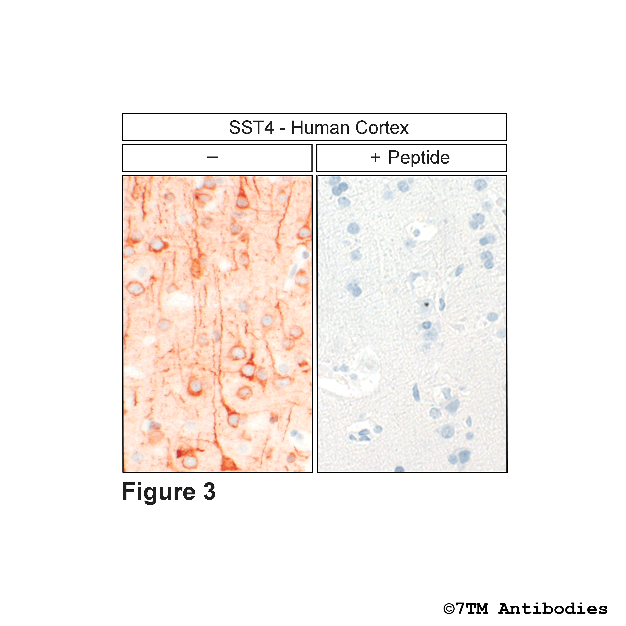
The non-phospho-SST4 receptor antibody is directed against the distal end of the carboxyl-terminal tail of human SST4. In can be used to detect total SST4 receptors in Western blots independent of phosphorylation. The non-phospho-SST4 antibody can also be used to isolate and enrich SST4 receptors from tissue and cell lysates. It also detects SST4 in cultured cells and tissue sections by immunohistochemistry.
| Alternative Names | SRIF2B |
| IUPHAR Target ID | 358 |
| UniProt ID | P31391 |
| Western Blot (WB) | 1:1000 |
| Immunocytochemistry (ICC) | 1:200 |
| Immunohistochemistry (IHC) | 1:100 |
| Species Reactivity | Human |
| Host / Isotype | Rabbit / IgG |
| Class | Monoclonal |
| Immunogen | A synthetic peptide presents the carboxyl-terminal tail of human SST4. |
| Form | Liquid |
| Purification | Antigen affinity chromatography |
| Storage buffer | Dulbecco's PBS, pH 7.4, with 150 mM NaCl, 0.02% sodium azide |
| Storage conditions | short-term 4°C, long-term -20°C |
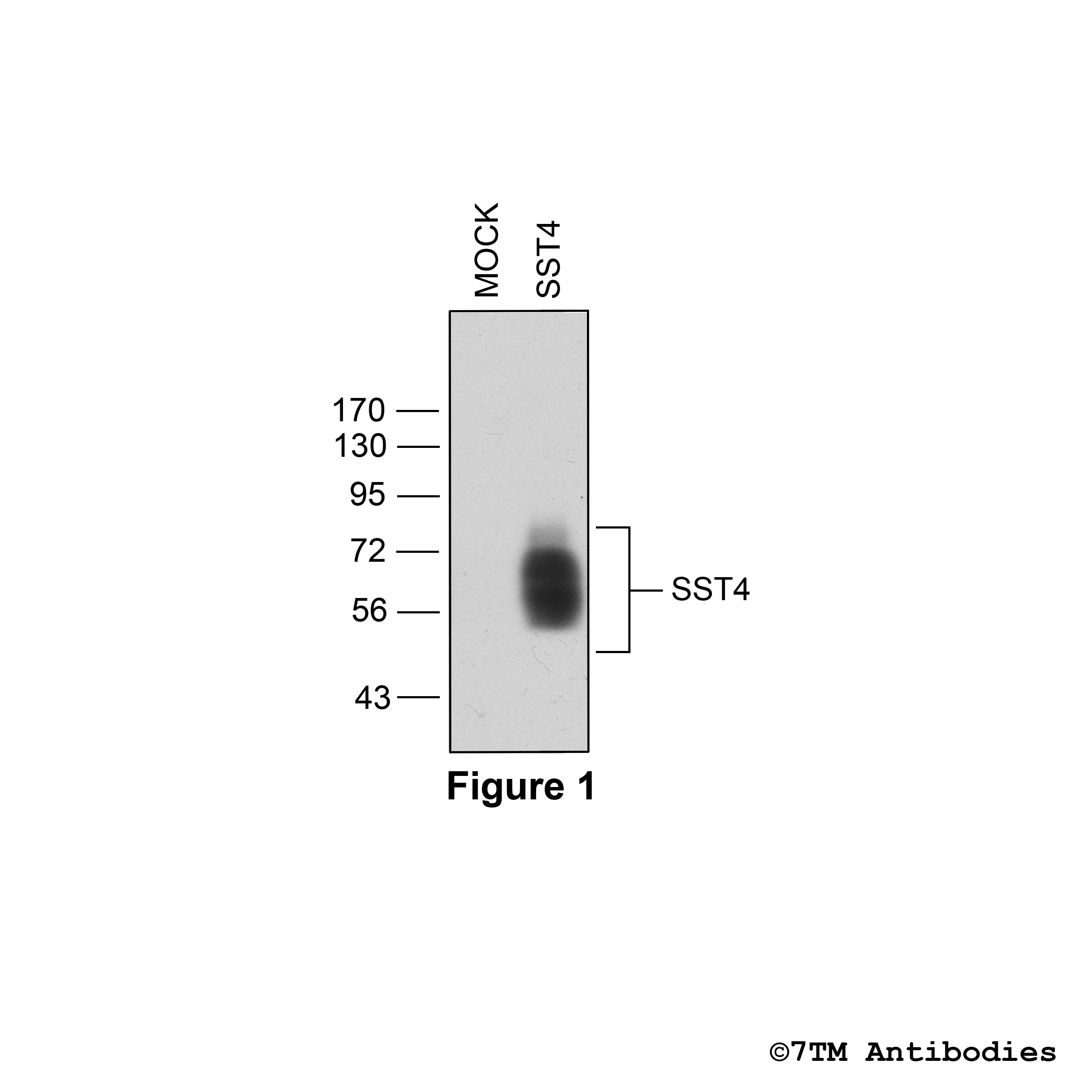
Figure 1. Validation of the Somatostatin Receptor 4 in transfected HEK293 cells. Native HEK293 cells (MOCK) or HEK293 cells stably expressing the Somatostatin Receptor 4 (SST4) were lysed and immunoblotted with the phosphorylation-independent anti-SST4 antibody (7TM0358N-RM) at a dilution of 1:1000.
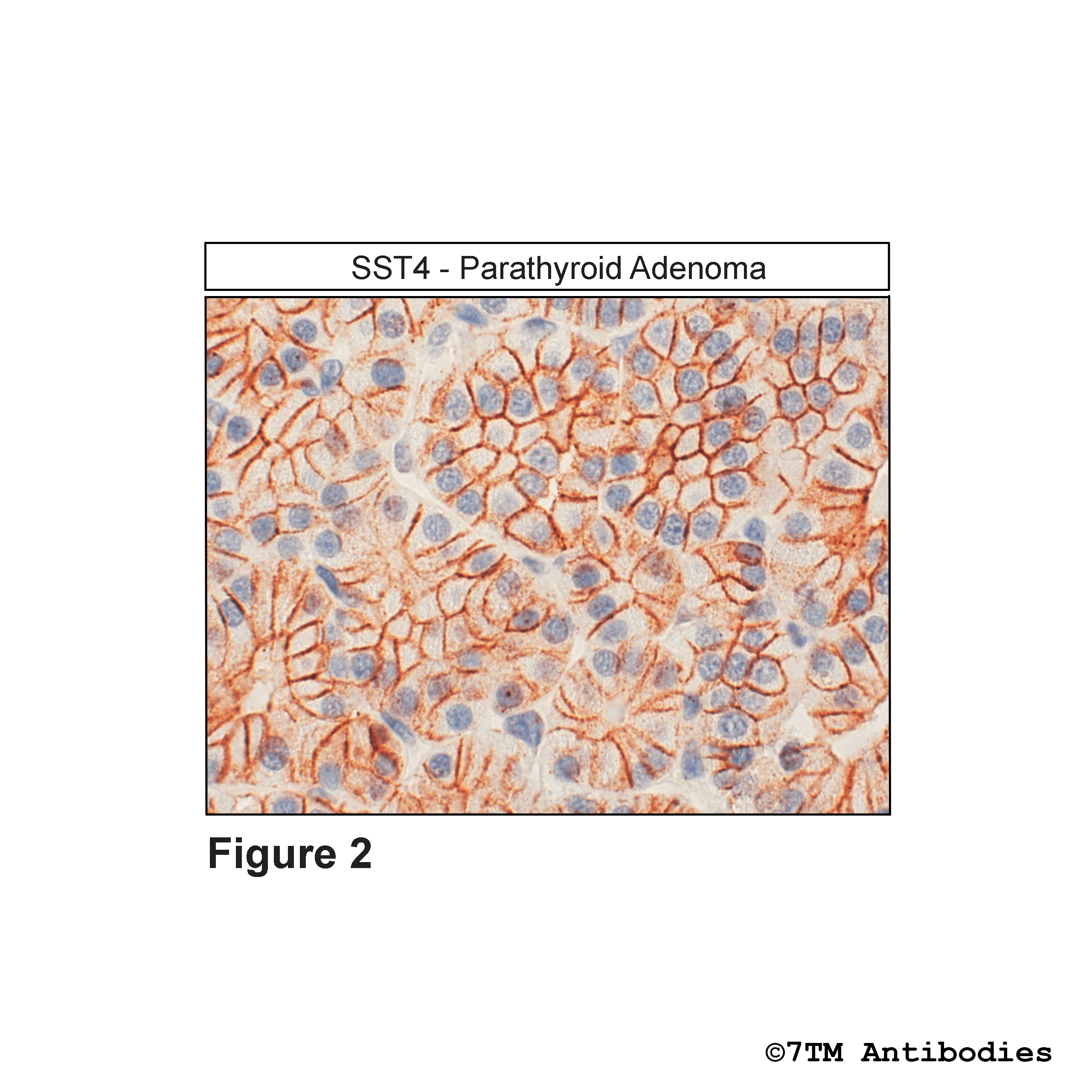
Figure 4. Immunohistochemical identification of Somatostatin Receptor 4 in parathyroid adenoma. Sections were dewaxed, microwaved in citric acid, and incubated with anti-SST4 (non-phospho-Somatostatin Receptor 4) antibody (7TM0358N-RM) at a dilution of 1:100. Sections were then sequentially treated with biotinylated anti-rabbit IgG and avidin-biotin solution.Color was developed by incubation in 3-amino-9-ethylcarbazole (ACE), and sections were counterstained with hematoxylin. Note, SST4 receptors were detected at the plasma membrane of nearly all cells in human parathyroid adenoma.
Figure 5. Immunohistochemical identification of Somastatin Receptor 5 in human anterior pituitary. Sections were dewaxed, microwaved in citric acid, and incubated with anti-SST5 (non-phospho-Somatostatin Receptor 5) antibody (7TM0359N-RM) at a dilution of 1:100. Sections were then sequentially treated with biotinylated anti-rabbit IgG and avidin-biotin solution.Color was developed by incubation in 3-amino-9-ethylcarbazole (ACE), and sections were counterstained with hematoxylin. Note, SST5 receptors were detected on a distinct population of cells in the anterior pituitary.
Figure 6. Immunohistochemical identification of Somastatin Receptor 5 in human growth-hormone-producing pituitary adenoma tissue. Sections were dewaxed, microwaved in citric acid, and incubated with anti-SST5 (non-phospho-Somatostatin Receptor 5) antibody (7TM0359N-ICRM) at a dilution of 1:100. Sections were then sequentially treated with biotinylated anti-rabbit IgG and avidin-biotin solution. Color was developed by incubation in 3-amino-9-ethylcarbazole (ACE), and sections were counterstained with hematoxylin. Note, SST5 receptors were uniformly detected at the plasma membrane of nearly all tumor cells.
Figure 7. Immunohistochemical identification of Somastatin Receptor 5 in human pheochromocytoma. Sections were dewaxed, microwaved in citric acid, and incubated with anti-SST5 (non-phospho-Somatostatin Receptor 5) antibody (7TM0359N-ICRM) at a dilution of 1:100. Sections were then sequentially treated with biotinylated anti-rabbit IgG and avidin-biotin solution.Color was developed by incubation in 3-amino-9-ethylcarbazole (ACE), and sections were counterstained with hematoxylin. Note, SST5 receptors were uniformly detected at the plasma membrane of nearly all cells in pheochromocytoma.